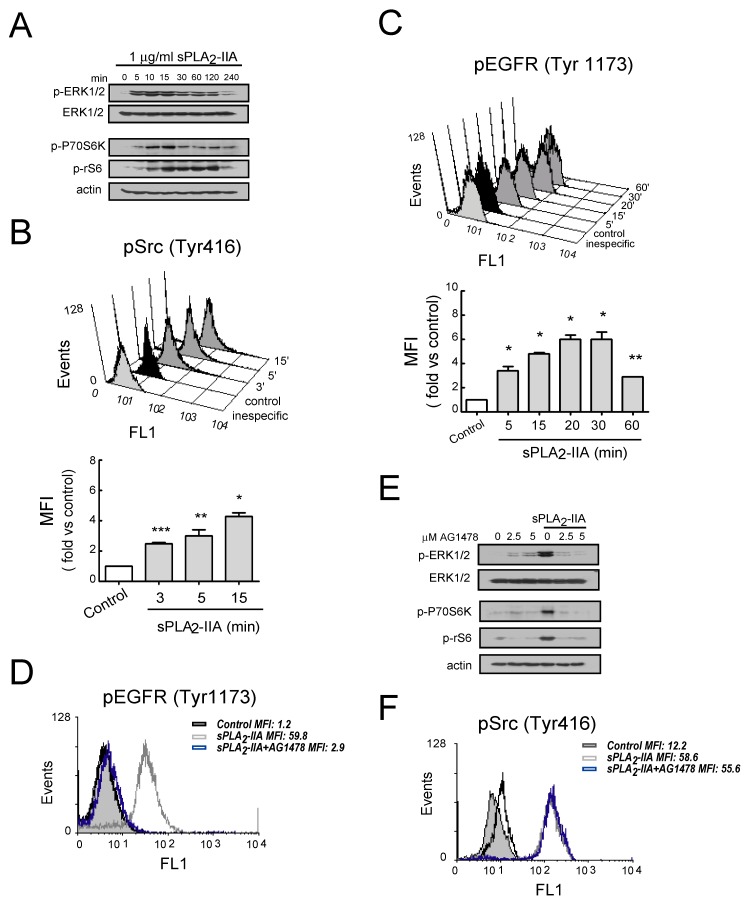
Figure 4.
sPLA2-IIA-activated intracellular signaling cascades in cardiac fibroblast (CFs) requires EGFR transactivation Cells were stimulated with 1 μg/mL of sPLA2-IIA for indicated times: using phospho-specific antibodies, (A) ERK, Akt, P70S6 and S6 phosphorylation were determined by immunoblotting, while (B) Src and (C) EGFR phosphorylation was examined by flow cytometry. Untreated (solid black curves) and sPLA2-IIA-treated (solid dark grey curves) CFs were compared with isotype controls (solid light grey curves). (D–F) Cells were pretreated with AG1478 before stimulation with sPLA2-IIA. (D) After 15 min, phospho-EGFR was studied by flow cytometry. (E) After 15 min, ERK, Akt, P70S6 and S6 phosphorylation were determined by immunoblotting, while (F) Src phosphorylation was studied by flow cytometry. Untreated (open black curves), sPLA2-IIA-treated (open grey curves), AG1478+sPLA2-IIA-treated (open blue curves) CFs were compared with isotype controls (solid light grey curves). * p < 0.001, ** p < 0.01 and *** p < 0.5 vs. control cells; mean ± SD; n = 3.

