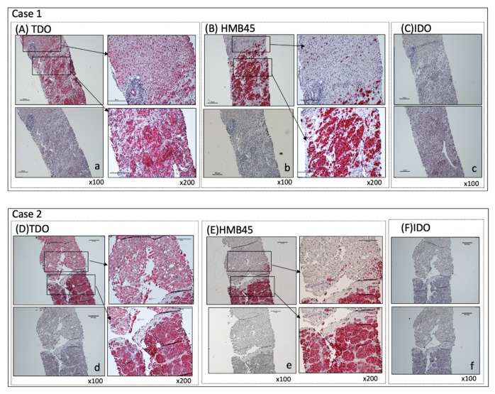
Figure 1.
Immunohistochemical (IHC) staining of liver metastasis specimens. Liver metastasis specimens were stained for TDO, HMB45, and IDO. Positive cells show a distinct red stain. IHC staining of two representative sets of archived liver metastasis specimens were shown. (A,D) TDO staining: For negative control, TDO antibody was blocked with specific blocking peptide (a,d). Magnification: ×100 (left panels); ×200 (right panels) Scale bar = 100 μm. (B,E) HMB45 staining: Melanoma metastasis is positive (right bottom) and surrounding liver tissue is negative (right top). Isotype-matched IgG was used as negative control (b,e). Magnification: ×100 (left panels); ×200 (right panels) Scale bar = 100 μm (C,F) IDO staining: IDO is negative for both metastatic melanoma and surrounding liver tissue (top). Isotype-matched IgG is used as negative control (c,f). Magnification: ×100. Scale bar = 100 μm.

