Figure 2.
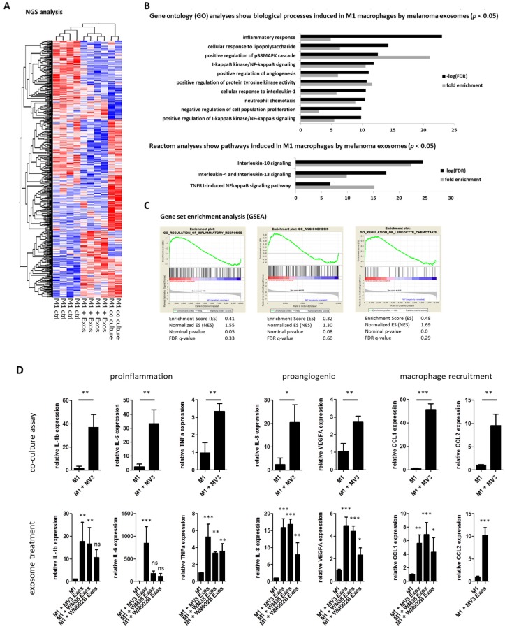
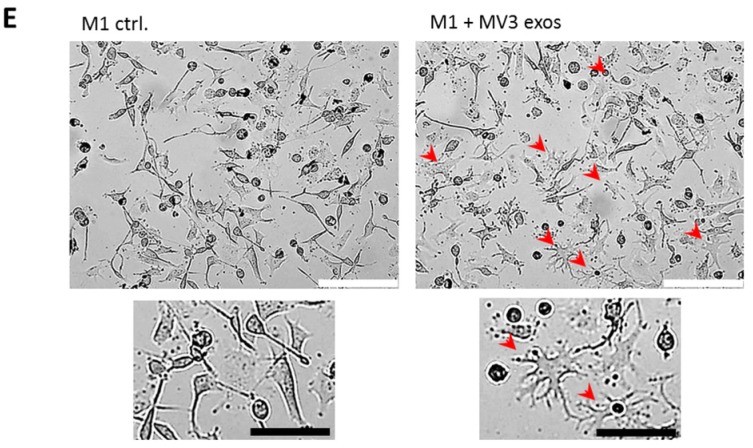
Melanoma exosomes induce a proinflammatory and proangiogenic phenotype macrophages. (A) Hierarchical clustering of differential transcripts of THP-1-derived M1 macrophages 48 h co-cultured with MV3 melanoma cells, treated with MV3 released exosomes or M1 macrophages alone. (B) Gene ontology and Reactome pathway analysis of genes significantly increased in M1 macrophages after treatment with melanoma-derived exosomes. (C) Gene set enrichment analysis (M1 + exosomes versus M1 control) of next-generation sequencing results shows induction of inflammation, angiogenesis, and leukocyte chemotaxis. (D) Quantitative RT-PCR validated induction of proinflammatory, proangiogenic, and chemotaxis-related gene expression after 48 h co-culture of M1 with MV3 cell line or treatment with melanoma cell-derived exosomes. Bars represent the average ± standard deviation of at least three independent experiments (* p ≤ 0.05; ** p ≤ 0.01; *** p ≤ 0.001; ns: not significant). (E) Bright-field microscopy shows representative morphological changes (red arrows) of THP-1-derived M1 macrophages with and without MV3- derived exosomes treatment (48 h). White scale bar: 200 µm; black scale bar: 50 µm.


