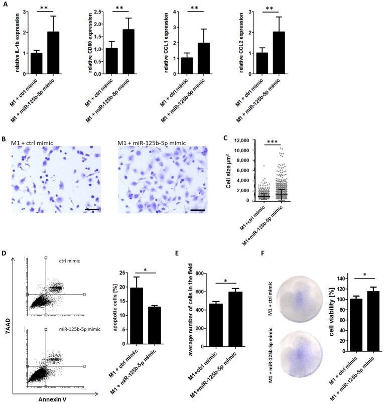
Figure 5.
Overexpression of miR-125b-5p partially mimics a exosome-induced TAM phenotype switch and promotes the survival of macrophages. THP-1-derived M1 macrophages were transfected for 48 h with miR-125b-5p mimics (100 nM) or a control mimic (100 nM), respectively. (A) qRT-PCR analyses show an induction of IL-1β, CD80, CCL1, and CCL2 after miR-125b-5p overexpression. Bars represent the average ± standard deviation of at least three independent experiments (** p ≤ 0.01). (B) Representative pictures of fixed and crystal violet-stained M1 macrophages show a morphological switch by miR-125b-5p overexpression. Scale bar: 100µm. (C) Quantification of the cell size of M1 macrophages 48 h after transfection with the control mimic or miR-125b-5p mimic (*** p ≤ 0.001). (D) Flow cytometry analysis of apoptosis 48 h after transfection. Bars represent the average ± standard deviation of at least three independent experiments (* p ≤ 0.05). (E) Cell count analysis of M1 polarized macrophages 48 h after transfection with the control mimic or miR-125b-5p mimic. Bars represent the average ± standard deviation of at least three independent experiments (* p ≤ 0.05). (F) Transfected macrophages were 72 h co-cultured indirectly with the MV3 melanoma cell line. Macrophages were fixed and stained with gentian violet viability stain. Representative pictures show stained M1 macrophages in a culture dish. Viability of macrophages was quantified measuring absorption at 620 nm by a multi plate reader. Bars show the average ± standard deviation of at least three independent experiments (* p ≤ 0.05).

