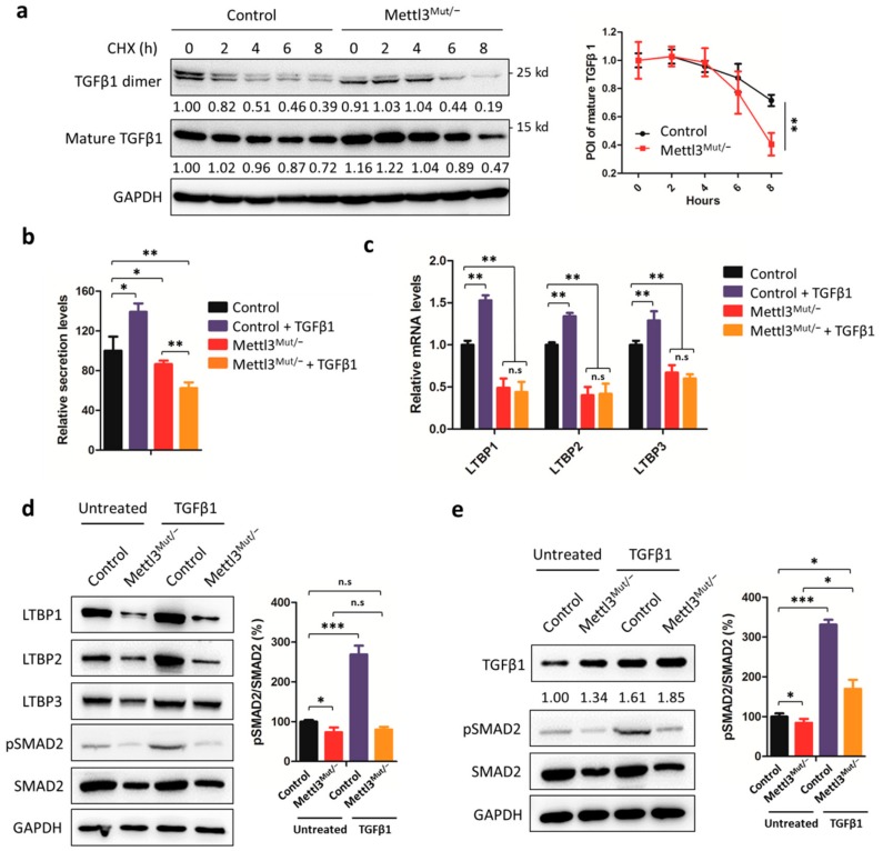
Figure 5.
Secretion of TGFβ1 is modulated by METTL3. (a) Control and Mettl3Mut/− HeLa cells were incubated with 100 µg/mL cycloheximide (CHX) for indicated times. Protein levels of mature TGFβ1 and TGFβ1 dimer were measured by Western blot (left). Band intensities were analyzed by ImageJ and are listed at the bottom of target bands. Mature TGFβ1 levels were quantitatively analyzed (right); (b) Control and Mettl3Mut/− HeLa cells were incubated with 10 ng/mL TGFβ1 for 48 h. Secretion of TGFβ1 in control and Mettl3Mut/− HeLa cells was measured by ELISA kit. The relative secretion levels of TGFβ1 were normalized to culture medium with or without the addition of TGFβ1; (c) Control and Mettl3Mut/− HeLa cells were incubated with 10 ng/mL TGFβ1 for 48 h. The expression levels of LTBP1, LTBP2, and LTBP3 mRNA in control and Mettl3Mut/− HeLa cells were measured by qRT-PCR; (d) Control and Mettl3Mut/− HeLa cells were incubated with 10 ng/mL TGFβ1 for 48 h. The expression levels of LTBP1, LTBP2, LTBP3, pSMAD2, and SMAD2 in control and Mettl3Mut/− HeLa cells were measured by Western blot (left). Percentages of pSMAD2 to SMAD2 were analyzed (right); (e) Control and Mettl3Mut/− HeLa cells were transiently overexpressed in LTBP1 for 24 h, then incubated with 10 ng/mL TGFβ1 for 48 h. The expression levels of TGFβ1, pSMAD2, and SMAD2 were measured by Western blot (left). Band intensities of TGFβ1 were analyzed by ImageJ and are listed at the bottom of targets. Percentages of pSMAD2 to SMAD2 were analyzed (right). Data are presented as means ± SD from three independent experiments. Student’s t-test, n.s, no significant; *, p < 0.05; **, p < 0.01; ***, p < 0.001 compared with control.

