Figure 6.
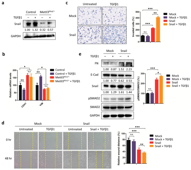
Snail is the core protein responsible for METTL3-mediated EMT. (a) Control and Mettl3Mut/− HeLa cells were incubated with 10 ng/mL TGFβ1 for 48 h. The expression levels of Snail were measured by Western blot. Band intensities of Snail were analyzed by ImageJ and are listed at the bottom of target bands; (b) Control and Mettl3Mut/− HeLa cells were incubated with 10 ng/mL TGFβ1 for 48 h. Expression levels of CDH1 and VIM mRNA were measured by qRT-PCR; (c) Mettl3Mut/− cells transiently overexpressing empty vector (Mock) and Snail were incubated with 10 ng/mL TGFβ1. Cells were allowed to invade for 24 h and were tested by CytoSelect™ 24-well Cell Invasion assay kits (8 µm, colorimetric format; left). Invaded cells were then quantitatively analyzed (right); (d) Mettl3Mut/− cells transiently overexpressing empty vector (Mock) and Snail were incubated with 10 ng/mL TGFβ1 for indicated times, and the wound healing of cells was recorded; scale bar, 100 µm; (e) Mettl3Mut/− cells transiently overexpressing empty vector (Mock) and Snail were incubated with 10 ng/mL TGFβ1 for 48 h. Expression levels of FN, E-Cad, Snail, pSMAD2, and SMAD2 were measured by Western blot (left). Band intensities of FN, E-Cad, and Snail were analyzed by ImageJ and listed at the bottom of targets. Percentages of pSMAD2 to SMAD2 were analyzed (right). Data are presented as means ± SD from three independent experiments. Student’s t-test, n.s, no significant; *, p < 0.05; **, p < 0.01; ***, p < 0.001 compared with control.

