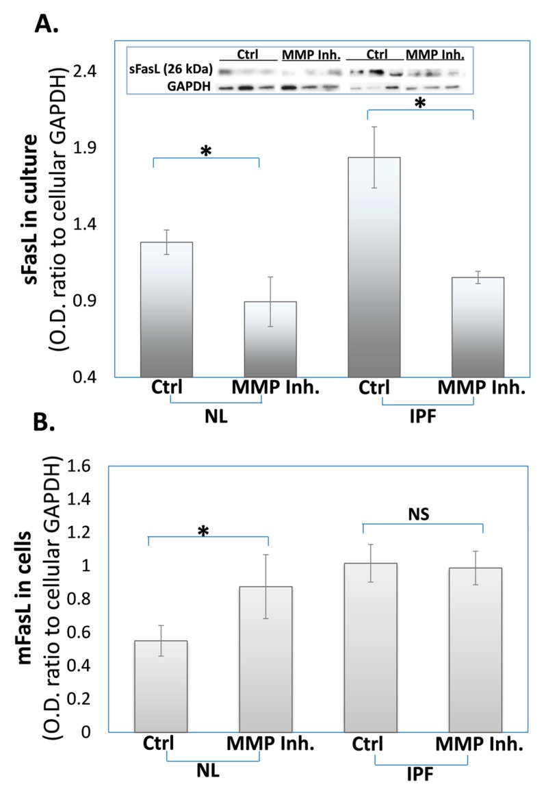
Figure 1.
The decreased soluble and increased membrane FasL levels in fibrotic-(IPF) and normal (NL)-lung myofibroblasts, following exposure to the batimastat MMP inhibitor. Western blot of: (A) sFasL in culture medium and (B) mFasL, of fibroblast cell lines (3 × 10⁵) of fibrotic-lung/ATCC191 (IPF)- or normal/ATCC151 (NL)-lungs; Graphical presentation and blots (insert) with optical density ratios normalized to fibroblasts GAPDH after treatment with control-vehicle (0.1% DMSO) or batimastat (24 h, 10 µM). Mean ± standard deviation; n = 4; * p < 0.05, NS—p > 0.05.

