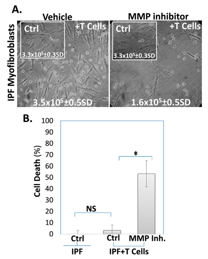
Figure 2.
The MMP inhibitor (batimastat) decreases IPF-lung myofibroblast’ resistance to T cell-induced cell death. (A) Light microscope images with trypan blue exclusion (inserted numbers); and (B) Graphical presentation of the cell death percentage defined by trypan blue exclusion. Comparisons were made between IPF-lung myofibroblasts (ATCC-191 cell-line) cultured alone (inserts-Ctrl) and IPF-lung myofibroblasts co-cultured with T cells (1 × 10⁶, 48 h), following being treated with vehicle (0.1% DMSO) or MMP-inhibitor (batimastat 10 µM, 24 h), (vehicle or MMP inhibitor, +T cells). The percentage of dead cells to total cell count in each sample was analyzed as the mean ± standard deviation; n = 3; * p < 0.05, NS p > 0.05.

