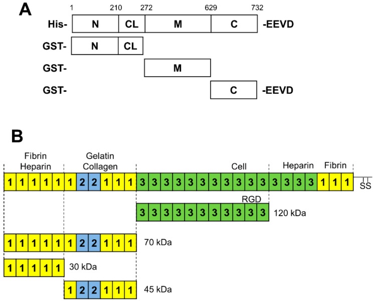
Figure 1.
Schematic diagram of HSP90 and fibronectin (FN) domains. (A) HSP90 domain boundaries indicated by numbering and recombinant fragments used in this study. (B) Domain structure of full-length fibronectin and proteolytic fragments thereof. The squares labeled 1, 2, and 3 refer to the type-I, type-II, and type-III FN domains, respectively. The binding sites of FN interactors are labeled above, while the sites of proteolytic cleavage of full-length FN are indicated by dotted lines and they give rise to the smaller 120, 70, 45, and 30 kDa fragments used in this study.

