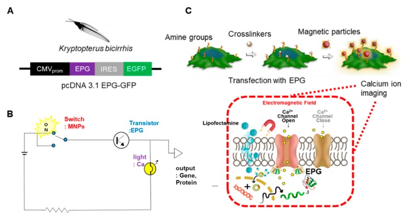
Figure 1.
Illustration of the remote regulation of [Ca2+]i in human embryonic kidney (HEKEPG+GFP) cells. (A) Plasmid encodes of both the electromagnetic perceptive gene (EPG) and the Green fluorescent protein (GFP) using Internal ribosome entry site (IRES) under the regulation of a general cytomegalovirus promotor (CMV promoter) were constructed. (B) Scheme of engineering biological circuit; switch = magnetic particles, transistor = EPG, light = Ca imaging, output = target gene/protein. (C) EPG+GFP was expressed in HEK293T. Sulfo-NHS-SS-Biotin was used to crosslink biotin with amine groups of cell-surface proteins. Streptavidin-coated Manetic particles (MPs) were added to the media and conjugated to biotin moieties on the cells (MPs are located outside of cells). [Ca2+]i imaging was employed to monitor the cell activation profiles with magnetic stimulation. EPG is a transmembrane protein; the origin of the Ca ion before the magnetic stimulation of cells is unknown; Ca ion is mainly stored in Endoplasmic reticulum (ER) but the uptake of Ca ion should be considered.

