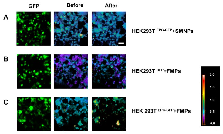
Figure 4.
Calcium ion imaging following stimulation with different types of magnetic particles. Images of [Ca2+]i in EPG+GFP-transfected HEK293T cells (the original GFP images: left column, 340/380 ratio pseudo-images before adding MPs: middle column, 340/380 nm ratio image after the addition of MPs: right column, 20×). (A) HEK293TEPG+GFP with SMNPs. (B) HEK293TGFP with Ferromagnetic particles (FMPs). (C) HEK293TEPG+GFP with FMPs.

