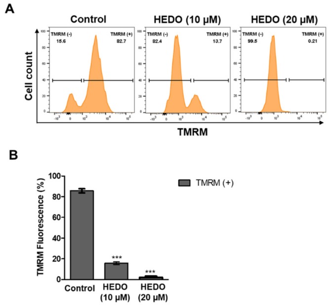
Figure 3.
Decrease in mitochondrial membrane potential in OCI-LY3 cells due to HEDO. (A) Mitochondrial membrane potential of OCI-LY3 cells loaded with tetramethylrhodamine methyl ester (TMRM) (100 nM), as detected by flow cytometry. (B) Quantification of the membrane potential in mitochondria, as measured by flow cytometry. Values indicate the means ± SEM. (n = 3, *** p ≤ 0.001).

