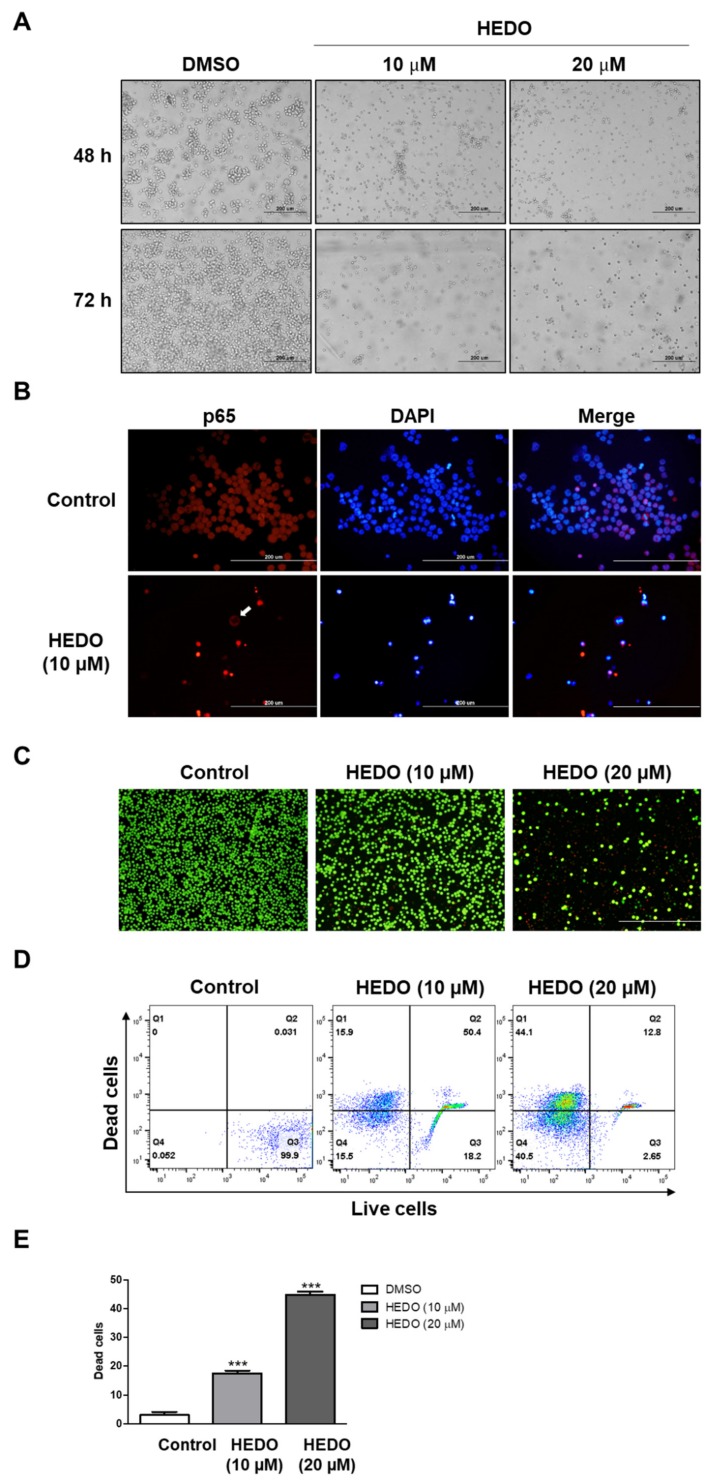
Figure 7.
Effects of HEDO on morphological changes, p65 nuclear translocation, and cell viability in OCI-LY3 cells. (A) Cells treated with HEDO (10 and 20 μM) show morphological changes that are characteristic of apoptosis at 48 and 72 h post-treatment using phase contrast microscopy (n = 3, 200x magnification, scale bar: 200 μm). (B) Effects of HEDO on p65 nuclear translocation in OCI-LY3 cells. OCI-LY3 cells were cultured in the presence or absence of HEDO (10 µM). The white arrows show that the nuclear translocation of p65 was inhibited by HEDO and the p65 remained in the cytoplasm. (C) Merged fluorescence images showing untreated and HEDO-exposed (48 h) OCI-LY3 cells (green fluorescence represents live cells and red fluorescence represents dead cells). (D) Flow cytometry analysis of live and dead Jurkat cells with calcein and ethidium homodimer-1 staining. (E) Quantification of dead cells. Values indicate the means ± SEM. (n = 3, *** p ≤ 0.001).

