Figure 1.
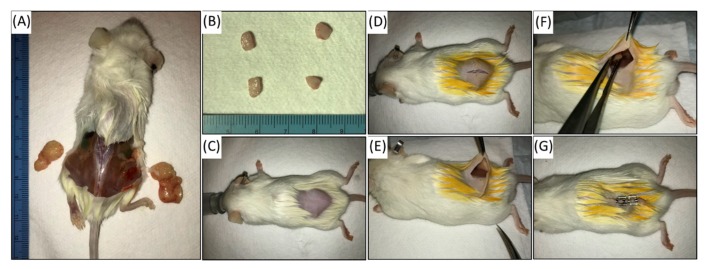
Passaging of patient-derived xenograft (PDX): NSG mice are euthanized once tumors reach approximately 1.5cm as measured by Vernier caliper (A). Tumors are explanted and divided into 5mm pieces for engraftment (B). Additional tumor specimen is collected at every passage for formalin fixation and paraffin embedding to confirm faithful maintenance of malignant histology. The new host NSG mouse at 6-8 weeks is anesthetized and prepped for grafting (C). The hind region is shaved and cleaned with alcohol and betadine. A 1cm midline incision allows for bilateral implantation, if desired (D,E). Tumor specimen is placed in subcutaneous pouch overlying the muscle (F). Matrigel can be used to keep tumor in position. Skin is closed, and mouse is started on amoxicillin for infection prophylaxis (G).

