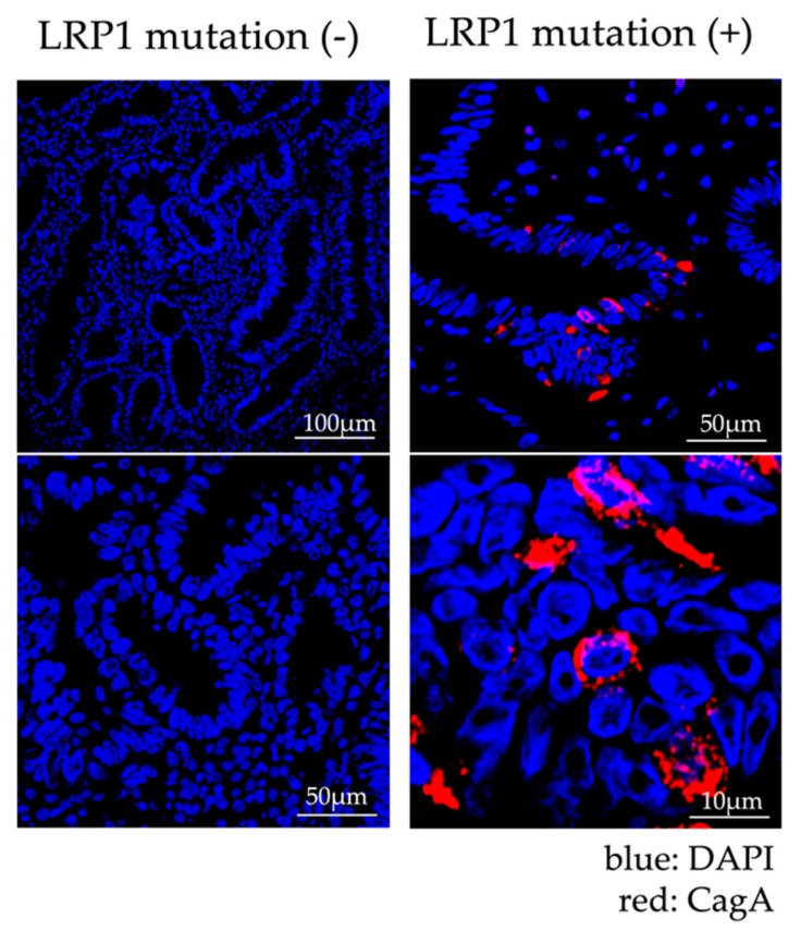
Figure 4.
Immunofluorescence Staining of specimens treated by endoscopic submucosal dissection (ESD). The case with LRP1 mutation is shown on the right. CagA staining (red) was observed in the cytoplasm of gastric cancer cells in LRP1 mutant cases. On the other hand, the case without LRP1 mutation is shown on the left. CagA staining was not found in the cytoplasm of gastric cancer cells in cases without LRP1 mutation. DAPI (4’,6-diamidino-2-phenylindole) nuclear staining is shown in blue.

