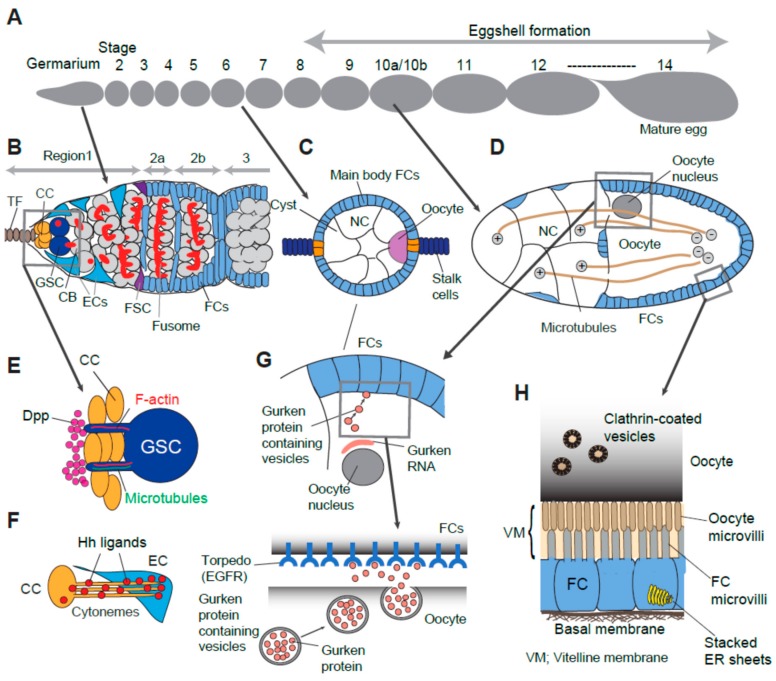
Figure 1.
Examples of usage of specialized structures and/or machineries for cell-cell interaction. (A) Overview of fourteen developmental stages of oogenesis. (B) Cellular organization of germarium. (C) Stage 6 egg chamber. Cyst contains 15 nurse cells and one posteriorly localized oocyte. Specified follicle cells (Polar, stalk and main body follicle cells) are determined by interplay of local signaling (See Section 4). (D) Stage 10b egg chamber. Nurse cells directly transfer their material (mRNA, organelle, protein) toward oocyte via microtubule tracks. Microtubule plus end are in nurse cells and their minus ends are in the oocyte. (E) A GSC with a cytocensor and an actin protrusion. Dpp ligands accumulate at the anterior face of CCs away from GSCs. (F) CC emanates cytonemes to deliver the Hh ligand toward EC. (G) Polarized Gurken secretion in a stage 10b egg chamber. The oocyte nucleus is positioned at the dorsal-anterior region of oocyte. Gurken mRNA are seen between the oocyte nucleus and oocyte-follicle cell interface. Polarized secretion of Gurken occurs locally at the dorso-anterior surface of oocyte and activates the Torpedo receptor expressed on the surface of nearby follicle cells. (H) Yolk material deposition from follicle cell into the oocyte (See details in Section 6). Oocyte microvilli and follicle cell microvilli extend and interdigitate each other between the space of the oocyte and follicle cell layer which is filled with vitelline membrane. CC (cap cells), TF (terminal filament), GSC (germline stem cell), CB (cystoblast), EC (escort cell), FSC (follicle stem cell), FCs (follicle cells), NC (nurse cell), Dpp (decapentaplegic), Hh (hedgehog), EGFR (epidermal growth factor receptor), VM (vitelline membrane).

