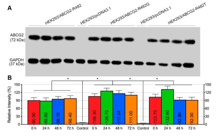
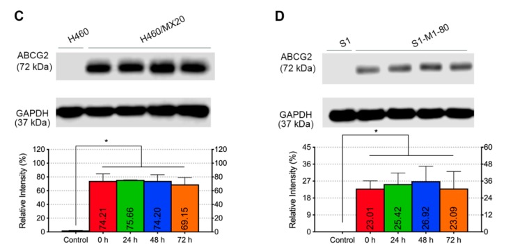
Figure 7.
Effect of venetoclax on the ABCG2 protein expression. (A) ABCG2 protein expression after incubation with 10 µM venetoclax at different time points in HEK293/pcDNA3.1, HEK293/ABCG2-R482, HEK293/ABCG2-R482G and HEK293/ABCG2-R482T cells. GAPDH was used as a loading control. (B) Expression level quantification by grey scale values. (C) ABCG2 protein expression after incubation with 10 µM venetoclax at different time points in H460 and H460/MX20 cells. (D) ABCG2 protein expression after incubation with 10 µM venetoclax at different time points in S1 and S1-M1-80 cells. Columns with error bars represent mean ± SD from 3 independent triplicate experiments. Asterisks (*) indicate p < 0.05 versus parental group (HEK293/pcDNA3.1).


