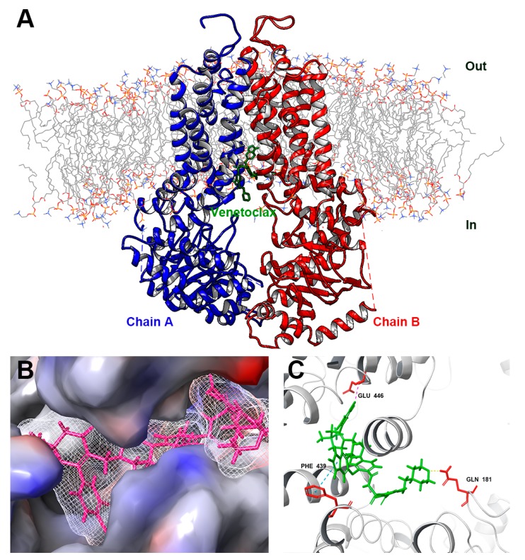
Figure 9.
Interaction between venetoclax and ABCG2 protein by docking simulation. (A) An overview of top-scored docked position. Venetoclax was colored green. Two chains showed as ribbons were colored red and blue. Lipid molecules in cytoplasm membrane were depicted as colored lines. (B) Interaction between venetoclax and ABCG2 binding pocket with molecular surface displayed. (C) Docked conformation of venetoclax and wild-type ABCG2. Venetoclax was colored green and important residues were colored red. π-π stacking was represented as cyan dotted line, hydrogen bond was represented as yellow dotted line, halogen bond was represented as magenta dotted line.

