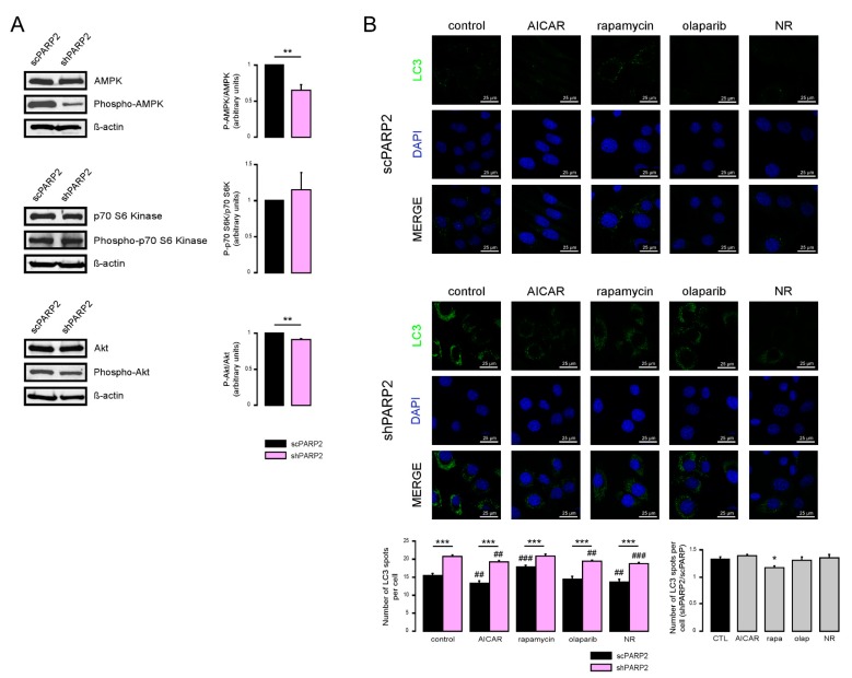
Figure 6.
Inhibition of AMPK and cellular NAD+ level modulate autophagy in a PARP2-dependent fashion. (A) In scPARP2 and shPARP2 C2C12 cells, the change of AMPK, phospho-AMPK, p70 S6 kinase, phospho-p70 S6 kinase, Akt, and phospho-Akt expression were analyzed by Western blotting (n = 3). (B) scPARP2 and shPARP2 C2C12 cells were treated with 1 mM AICAR, 20 nM rapamycin (rapa), 1 µM olaparib (olap), or 500 µM nicotinamide-riboside (NR) for 24 h. LC3 expression was assessed in confocal microscopy experiments. Alexa Fluor 488-linked LC3 specific antibody was used and the nuclei were visualized using DAPI and vesicles were counted. Representative images are presented in the figure. ## and ### represent statistically significant differences between the control and treated cells at p < 0.01 and p < 0.001, respectively. *, ** and *** represent statistically significant differences between the scPARP2 and shPARP2 cells at p < 0.05, p < 0.01 and p < 0.001. For the determination of statistical significance, ANOVA test was used followed by Tukey’s post hoc test.

