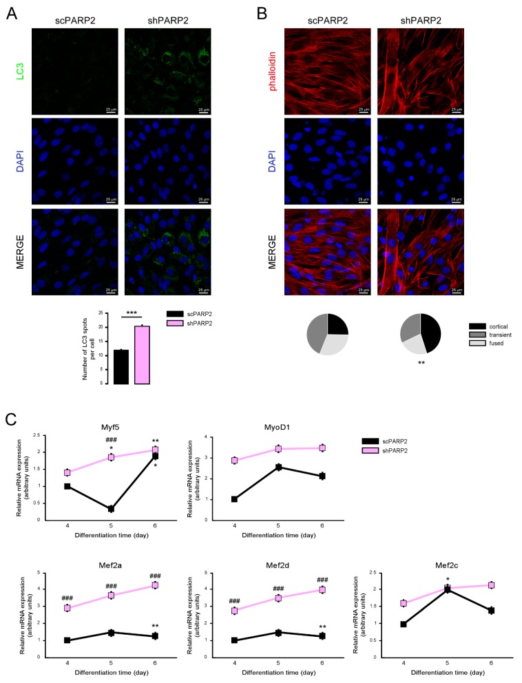
Figure 12.
Silencing of PARP2 supports myogenic differentiation. A total of 20,000 scPARP2 and shPARP2 C2C12 cells were seeded in 24-well plates that were differentiated for 4 days then (A) LC3-positive vesicles were assessed by immunofluorescence, (B) actin cytoskeleton was stained by Texas Red-X Phalloidin+DAPI, and (C) the expression of a set of myogenic genes were assessed by RT-qPCR. On panel B 100/100 cells were scored for the structure of actin cytoskeleton as diffuse, transient, and strong cortical staining. *** represent statistical difference between scPARP2 and shPARP2 cells at p < 0.001 on panel A. ** represent statistical difference between scPARP2 and shPARP2 cells in terms of actin morphology at p < 0.01 on panel B. In panel C, ### represents statistically significant differences between the scPARP2 and shPARP2 cells at p < 0.001. * and ** represent statistically significant differences between the control and differentiated cells at p < 0.05 and p < 0.01, respectively. For the determination of statistical significance, ANOVA test was used followed by Tukey’s post hoc test on panels A and C, while on panel B chi square test was applied.

