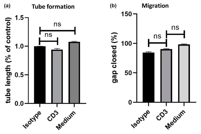
Figure 3.
human umbilical vein endothelial cell (HUVECs) were exposed to anti-CD3 treated PBMCs. Cell migration and angiogenesis were assessed by (a) tube assay and (b) tube formation, respectively. Compared to isotype control (100%), exposure to supernatant of anti-CD3 treated PBMC did not induce (a) endothelial cell tube formation (94 ± 2%; p = n.s. versus isotype control) (b) nor chemotaxis (90 ± 1%; p = n.s.) in HUVECs.

