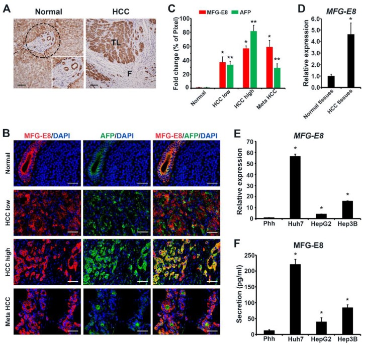
Figure 1.
Expression of milk fat globule-EGF factor 8 (MFG-E8)in clinical hepatocellular carcinoma (HCC) tissues and HCC cell lines. (A) Immunohistochemical staining of MFG-E8 in HCC tissues and normal liver tissues obtained from patients with HCC. Inset, enlarged image of the dashed circular area. TL, tumor lobule; F, fibrous septa. Scale bars, 100 μm. (B) Representative immunofluorescence images of primary and metastatic HCC tissues, showing the expression of MFG-E8 and AFP. Cell nuclei were stained with DAPI. Scale bars, 50 μm. Quantitation of immunoreactive cells are shown in (C). The percentages of positive immunofluorescence signal intensities of total image (% of pixel) were measured using ImageJ software and expressed as relative values to those in normal tissues. (D,E) The mRNA expression levels of MFG-E8 in normal tissues and HCC tissues (D) and in human primary hepatocytes (Phh) and HCC cell lines (Huh7, HepG2, and Hep3B) (E). mRNA expression data were normalized to GAPDH levels and expressed as relative values. (F) The levels of MFG-E8 protein secreted from human Phh and HCC cells. Data represent the mean ± S.D. * p < 0.05 versus normal tissue (C,D) and Phh (E,F) by a two-tailed Student’s t-test.

