Figure 9.
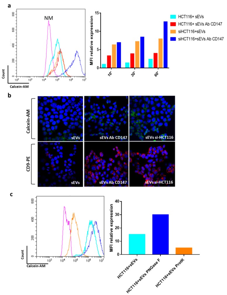
Blocking CD147 ON sEVs increases their cellular internalization. (a) Analysis of sEVs (small extracellular vesicles) uptake in recipient HCT116 and in CD147 knockdown HCT116 cells (siHCT116) compared to the uptake of sEVs blocked with CD147 antibody (sEVs CD147); data representative of three independent experiments are shown by bar charts on the right; (b) qualitative confirmation of Calcein-AM or anti-CD9 antibodies stained sEVs uptake in control HCT116 and in CD147 knockdown HCT116 cells (si-HCT116) by confocal microscopy. DAPI (blue) = staining nuclei; Calcein-AM = green; CD9-PE = red. (c) Same experiment as described in (a) using sEVs pretreated with proteinase K (ProtK) or N-Glycosidase F (PNGase F). Representative examples of staining after 1hours of incubation with CR-CSC–released sEVs. The images are representative of three independent experiments. NM = HCT116 treated for 1 h with Re121-sEVs not-stained with 1 μM Calcein-AM; HCT116 sEVs = HCT116 recipient cells treated for 1 h with sEVs stained with 1 μM Calcein-AM; siHCT116 + sEVs = HCT116 recipient cells transfected with non-targeting control and treated for 1 h with Re121 sEVs stained with 1 μM Calcein-AM; HCT116 + sEVs Ab CD147 = HCT116 cells treated with Re121 sEVs CD147 blocked with antibody. Magnification= 600×.

