Figure 1.
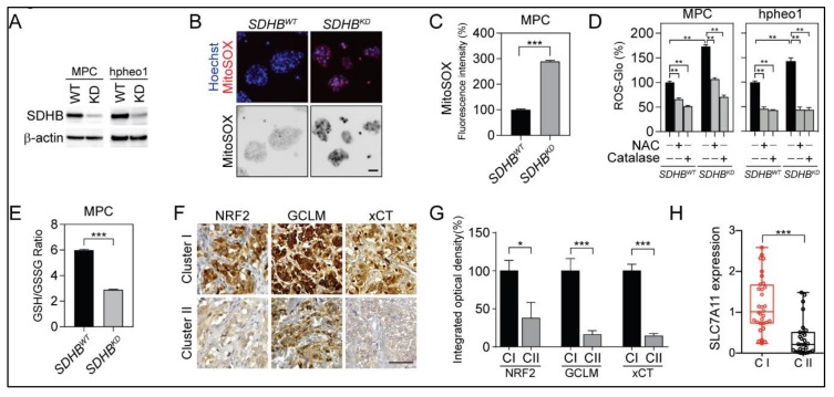
Succinate dehydrogenase subunit B (SDHB) deficiency reprogrammed reactive oxygen species (ROS) homeostasis and glutathione metabolism. (A) Immunoblotting showed the expression levels of SDHB in mouse pheochromocytoma (MPC) and hpheo1 cells. β-actin was used as internal control. (B) MitoSOX-Red staining showed ROS accumulation in SDHB knock down (SDHBKD) MPC cells. Bar = 10 μm. (C) Flow cytometry analysis showed elevated MitoSOX-Red staining in SDHBKD compared to SDHB wild type (SDHBWT) MPC cells. *** p < 0.001. (D) ROS quantification showed increased ROS in SDHBKD MPC and hpheo1 cells. Exogenous ROS scavenger N-acetylcysteine (NAC) and catalase reduced ROS level. ROS signal was measured and normalized to protein quantification. ** p < 0.01. (E) Glutathione quantification showed reduction of glutathione/glutathione disulfide (GSH/GSSG) ratio in SDHBKD compared to SDHBWT MPC cells. *** p < 0.001. (F) Immunohistochemistry staining showed that nuclear factor erythroid 2-related factor 2 (NRF2), glutamate-cysteine ligase regulatory subunit (GCLM), and cystine/glutamate transporter (SLC7A11, xCT) were increased in cluster I pheochromocytomas and paragangliomas (PCPGs). Bar = 50 μm. (G) Integrated optical density quantification for results shown in Figure 1F. For cluster I (CI), n = 4; for cluster II (CII), n = 4. * p < 0.05; *** p < 0.001. (H) Quantitative real-time PCR showed that SLC7A11 messenger RNA (mRNA) was increased in cluster I (C1; n = 8) compared to cluster II (CII; n = 7) PCPG specimen. *** p < 0.001.

