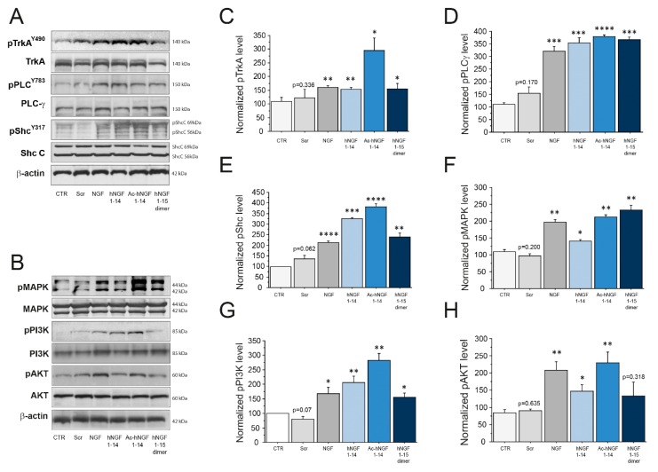
Figure 1.
Activation of the NGF-TrkA signalling pathway in cholinergic neurons by hNGF1–14 peptides. (A, C–E) Representative western blotting (A) and densitometric analyses (C–E) of pTrkA (A,C), and early Trk signalling adaptors pPLC-γ (A,D), pShc (A,E) in cholinergic neurons treated for 7′ with 10 μM scrambled hNGF1–14 (Scr), 10 μM hNGF1–14, 10 μM Ac-hNGF1–14, 10 μM hNGF1-15 dimer and 100 ng/mL (3.84 nM) NGF. (B, F–H). Representative western blotting (B) and densitometric analyses (F–H) of pMAPK (B,F), pPI3K (B,G), pAKT (B,H) in cholinergic neurons treated for 15-20′ with 10 μM scrambled hNGF1–14 (Scr), 10 μM hNGF1–14, 10 μM Ac-hNGF1–14, 10 μM hNGF1–15 dimer and 100 ng/mL (3.84 nM) NGF. The phosphorylated level of each signalling molecule was reported as ratio over the corresponding total protein, and further normalized using β-actin as loading control. Data from n = 3 independent experiments were expressed as percentage of CTR and reported as mean +SEM. * p < 0.05; ** p < 0.01; *** p < 0.001; **** p < 0.0001. Full-length blots are presented in the Additional file.

