Figure 2.
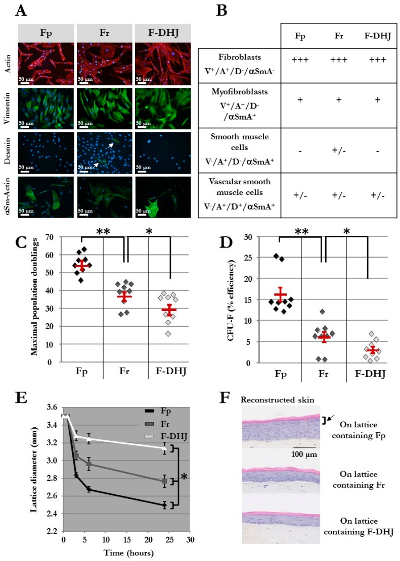
Phenotypic and functional properties of Fp, Fr, and F-DHJ fibroblasts. (A) Detection of actin (ACT) and vimentin (VIM), desmin (DES), and α-smooth muscle actin (α‑SMA) by immunochemistry in cultured Fp, Fr, and F-DHJ fibroblasts in the perspective of scoring according to Gabbiani’s classification [24]. White arrow points to rare DES+ cells present within the Fr population. (B) Scoring of Fp, Fr, and F‑DHJ fibroblasts according to ACT, VIM, DES, and α‑SMA detection: (−) = not present, (+/−) = low representation, (++) = frequent representation, and (+++) = major representation. (C) Long-term growth capacity of Fp, Fr, and F‑DHJ cells. Maximal cumulative population doubling values obtained with samples from independent donors are shown. Means ± SEM are indicated (* p < 0.05, ** p < 0.01; Wilcoxon test). (D) Colony-forming unit efficiency of Fp, Fr, and F‑DHJ cells. Fibroblast colony-forming unit (CFU-F) efficiency values (% of plated cells) obtained with samples from independent donors are shown. Means ± SEM are indicated (* p < 0.05, ** p < 0.01; Wilcoxon test). (E) Contractile capacity of Fp, Fr, and F‑DHJ cells in the 3D context of collagen lattices. Kinetics of lattice diameter evolutions. Means ± SEM are indicated (values obtained with samples from 9 independent donors) (* p < 0.05, Friedman’s test). (F) Efficiency of Fp, Fr, and F‑DHJ cells in promoting epidermis organogenesis by keratinocytes in a 3D reconstructed skin model. Representative reconstructed skin sections are shown (3 independent donors, each fibroblast sample tested in triplicates). The black arrow points to the epidermal granular layer that was obtained only in the presence of Fp fibroblasts.

