Figure 8.
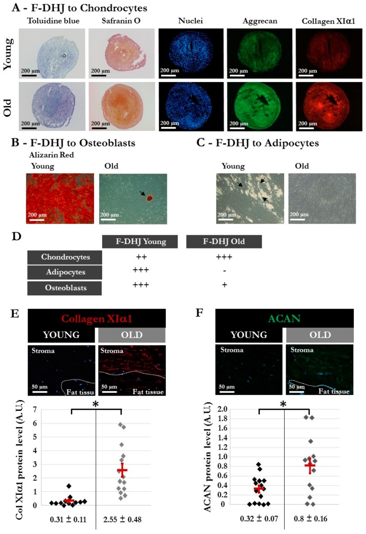
Different characteristics of DHJ components in skin from “young” and “old” donors. The “young” group comprised 5 donors (20, 22, 25, 28, and 31 years old) (same donors as in Figure 3), and the “old” group comprised 4 donors (55, 61, 65, and 65 years old). (A–C) Capacity of “young” and “old” F-DHJ cells to differentiate in vitro into three mesenchymal lineages: (A) chondrocytes (toluidine blue and safranin O staining, aggrecan (ACAN) and collagen XIα1 (ColXIα1) expression); (B) adipocytes (presence of cytoplasmic lipid droplets, black arrows); and (C) osteoblasts (alizarin-red staining). For panels (A–C), representative photographs are shown. (D) Summary of the differentiation capacity into chondrocytes, adipocytes, and osteoblasts of F-DHJ from “young” and “old” skin. Scoring of differentiation capabilities are presented: (−) = not present, (+) = low representation, (++) = frequent representation, and (+++) = major representation. (E,F) Immunofluorescence detection of ColXIα1 (E) and ACAN (F) in skin biopsies from “young” and “old” donors. Representative photographs are shown, in association with quantification values corresponding to a total of 12 (E) and 15 (F) regions of interest (ROI) for the 3 analyzed “young” donors (20, 22, and 28 years old) and a total of 14 (E) and 15 (F) ROI for the 3 analyzed “old” donors (57, 61, and 65 years old). Means ± SEM are indicated (* p < 0.05, Wilcoxon test).

