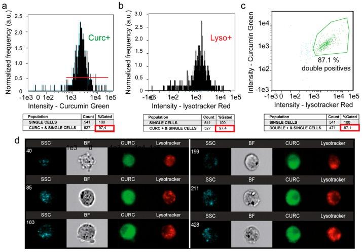
Figure 4.
Image cytometry (Amnis) of the double staining: LysoTracker Red and curcumin. (a) Curcumin green fluorescence for 24-h incubation with 20 µM curcumin. (b) LysoTracker Red fluorescence analysis for 10-min incubation with 100 nM LysoTracker Red. (c) Fluorescence histogram of curcumin and LysoTracker green fluorescence. (d) A selection of 6 cells analyzed with Amnis where it is possible to see the widespread intracellular staining of 5 µM curcumin for 3 h and the punctate staining with LysoTracker Red.

