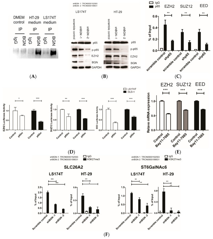
Figure 3.
Suppression of BGN inactivates NF-κB and decreases PRC2 activity in the promoter regions of SLC26A2 and ST6GalNAc6. (A) Western blot experiments of BGN in HT-29 and LS174T cultured medium. DMEM medium serves as a negative control. (B) Western blot experiments of phosphorylated p65 (p-p65) in LS174T and HT-29 after the introduction of shRNA for BGN. (C) ChIP/RT-qPCR results of p65 levels in EZH2, SUZ12, and EED regulatory regions in DLD-1 cells. (D) Mutational analysis of the p65-binding site in EZH2 p(+994/+841 in pGL3 promoter vector), SUZ12 p(−231/+69 in pGL3 basic vector), and EED p(−348/−49 in pGL3 basic vector). The mutated p65 construct (p65m) had a mutation of p65 binding site at bases +885 to +876 (from CCCCTAAAGC to TTTTTTTTT) in EZH2 and at bases −114 to −105 (from GGGGAATCCGC to AAAAAATAAAA) in SUZ12 and a mutation of p65 binding site at bases −219 to −210 (from GGGTACTTTCCC to AATACAAAAA) in EED. The control and mutated p65 constructs were transiently expressed in DLD-1 and LS174T cells for reporter assays. ***, p < 0.001;**, p < 0.01;*, p < 0.05 (n = 3 with mean ± SD shown). (E) EZH2, SUZ12, and EED mRNA in LS174T cells after administration with Bay11-7085 (10 μg/ml) for 6 h were analyzed by RT-qPCR. (F) ChIP/RT-qPCR results of H3K27me3 level in SLC26A2 and ST6GalNAc6 promoter regions in LS174T and HT-29 cells.

