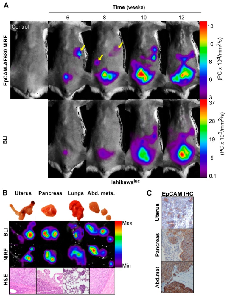
Figure 3.
Optical imaging of tumor growth in an orthotopic Ishikawaluc+ xenograft model. In vivo BLI and EpCAM-AF680 NIRF imaging of primary tumor growth in mice orthotopically implanted with Ishikawaluc+ cells. Metastatic lesions (arrows) were detected at an earlier time point in EpCAM-AF680 NIRF images. A tumor free mouse was used as control (upper left) (A). Macroscopic images of uterus, pancreas, lungs and abdominal metastases (B, upper panel) and corresponding ex vivo BLI and EpCAM-AF680 NIRF images (B, middle panels). Tumor cells are demonstrated in H&E stained sections (20x magnification) (B, lower panel). Positive EpCAM expression in primary tumor and metastases is demonstrated by IHC (C). Abbreviations: Bioluminescent imaging (BLI), Epithelial cell adhesion molecule (EpCAM), Hematoxylin and eosin (H&E), Immunohistochemistry (IHC), and Near-infrared fluorescence (NIRF).

