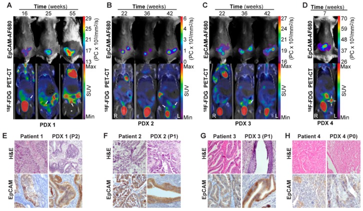
Figure 5.
In vivo imaging of tumor growth in PDX models using EpCAM-AF680 NIRF and 18F-FDG PET/CT. Longitudinal monitoring of uterine tumors of different histologic types in PDX models using EpCAM-AF680 NIRF and 18F-FDG PET/CT imaging. Arrows mark probable uterine tumors in PET/CT images (A–D). H&E staining demonstrating uterine tumor cells, and positive EpCAM IHC staining of uterine tumors from both donor patients and mouse xenografts (20× magnification) (E–H). Large bladder removed from image for visualization purposes. An uncropped version of this image can be found in Figure S2. Abbreviations: Epithelial cell adhesion molecule (EpCAM), Fluorine-18-fluorodeoxyglucose (18FDG), Hematoxylin and eosin (H&E), Near-infrared fluorescence (NIRF), Patient-derived xenograft (PDX), Positron emission tomography/computed tomography (PET/CT), and Standardized uptake value (SUV).

