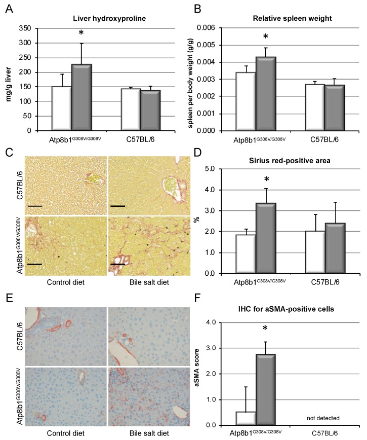
Figure 3.
Chronic cholestasis in Atp8b1G308V/G308V mice induces liver fibrosis in the presence of GCDCA. Atp8b1G308V/G308V and wild-type mice (C57BL/6) were fed a standard diet (white bars) or a CA (0.1% w/w)- plus GCDCA (0.3% w/w)-enriched diet (grey bars) to induce cholestasis and a humanized bile salt pool for 8 weeks. Liver hydroxyproline was determined as described (A). Spleen weight was determined at sacrifice and is presented relative to body weight (B). Representative Sirius Red staining of liver tissue is shown (C, black bar represents 50 µm) and Sirius-Red-positive area was quantified (D). Results are shown as mean ± standard deviation (n = 4 for C57BL/6 and n = 7 for Atp8b1G308V/G308V, * p < 0.05, t-test). IHC for αSMA was performed and representative slides are shown (E). Blinded histological scoring for αSMA was performed (F) and is expressed as mean ± standard deviation (n = 4, * p < 0.05, Pearson’s chi-squared test).

