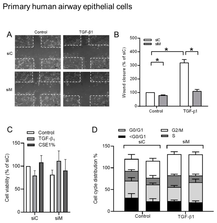
Figure 9.
The role of Ezrin, AKAP95, and Yotiao in TGF-β1-induced cell migration while using pHAE cells. (A) Representative images of wound healing assay after 24 h post scratch. The white dotted line indicated borders of scratches at 0 h. (B) Quantification of wound closure of TGF-β1 treated cells in co-cultured with combined knockdown of Ezrin, AKAP95 and Yotiao. (C) MTT assay in pHAE cells that were transfected with a combination siRNA of Ezrin, AKAP95, and Yotiao together with 3 ng/mL TGF-β1. (D) Cell cycle distribution in pHAE cells transfected with a combination siRNA of Ezrin, AKAP95 and Yotiao together with 3 ng/mL TGF-β1. Data represent 3–5 independent experiments. The data are expressed as mean ± SEM, * p < 0.05; significant difference between indicated groups.

