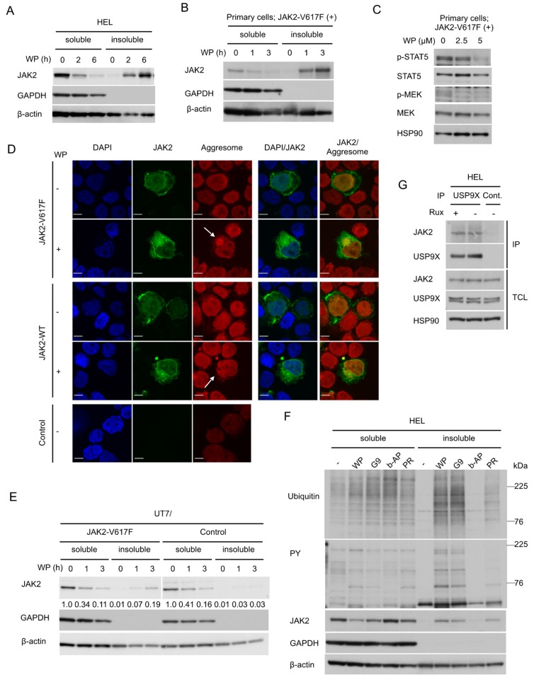
Figure 3.
WP1130 induces aggresomal translocation of JAK2 preferentially for the V617F mutant most likely through inhibition of USP9X. (A,B) HEL (A) or primary post-myeloproliferative neoplasms (MPN) secondary acute myeloid leukemic (sAML) cells expressing JAK2-V617F (B) were treated with 5 μM WP1130 (WP) for indicated times. Cells were lysed and detergent-soluble and –insoluble proteins were extracted and analyzed. GAPDH and β-actin were used for loading controls and confirmation of appropriate fractionation. (C) Primary post-MPN sAML cells expressing JAK2-V617F were treated for 2 h with indicated concentrations of WP1130 and analyzed. HSP90 was used for a loading control. (D) 293T cells were transfected with plasmids coding for JAK2-V617F, -WT, or empty vector (Control). Cells were left untreated as control or treated with 5 μM WP1130 for 3 h, followed by processing for confocal microscopy as described in the Materials and Methods. Images represent a 60× optical zoom with a 3× digital zoom. DAPI staining shows the position of the nucleus. Representative images of cells are shown. Positions of aggresomes are indicated by arrows. (E) UT7/JAK2-V617F or vector-control cells (Control) were treated with 5 μM WP1130 for indicated times and analyzed. Relative levels of JAK2 as compared with that in cells not treated with WP1130 were determined by densitometric analyses. (F) HEL cells were left untreated as control or treated for 3 h with 5 μM WP1130, 3 μM G9, 1 μM b-AP15, or 25 μM PR-619, as indicated. Cells were lysed and detergent-soluble and -insoluble protein were extracted and analyzed. (G) HEL cells were treated for 4 h with or without 2 μM ruxolitinib (Rux), as indicated. Immunoprecipitates (IP) with anti-USP9X antibody or normal rabbit IgG (Cont.) and total cell lysate (TCL) were analyzed.

