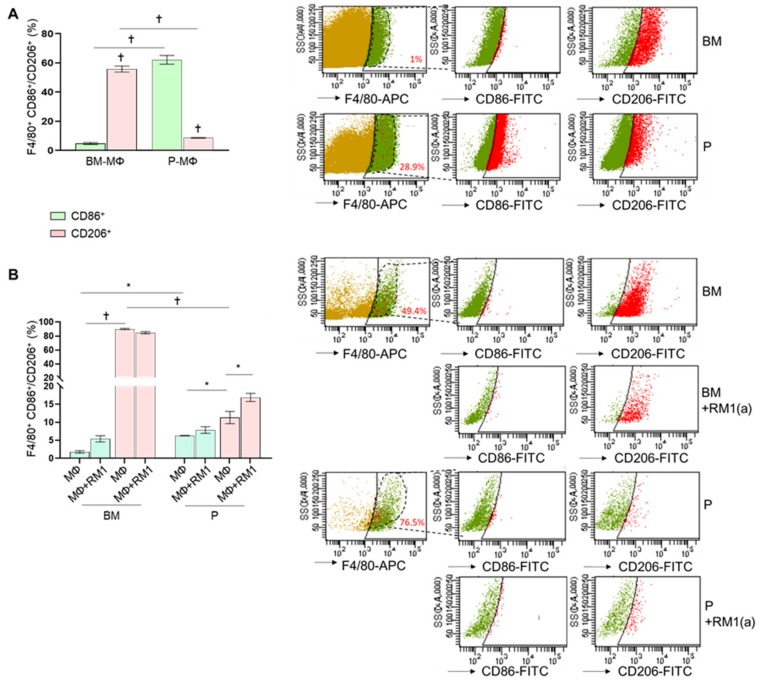
Figure 2.
Ex vivo and in vitro M1 and M2 polarization of bone marrow-derived and peritoneal macrophages. BM-MΦs and P-MΦs were isolated from C57BL/6J mice. (A) Freshly isolated bone marrow and peritoneal exudate cells were stained for anti-F4/80-APC combined with either anti-CD86-FITC or anti-CD206-FITC and analyzed by flow cytometry. (B) BM-MΦs and P-MΦs co-cultured with RM1(a) cells for 18 h were stained with anti-F4/80-APC combined with either anti-CD86-FITC or anti-CD206-FITC and analyzed by flow cytometry. BM, bone marrow; P, peritoneal. Data in A and B are mean ± SEM, n = 3 per group; *p < 0.05, †p < 0.0001 (one-way ANOVA; Dunnet’s multiple-comparisons test).

