Figure 2.
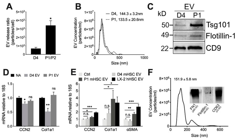
Characterization of EVs from mouse HSC. NTA was performed on EVs that had been purified by differential centrifugation of 2-day serum-free conditioned medium from D4 HSC or P1-P2 HSC, with particle number expressed as a function of (A) cell number or (B) particle size (mean ± S.E.M. for particle diameter (nm) is indicated). (C) Western blot analysis of D4 or P1 EVs (25 µg EV protein per lane) showing the presence of common EV proteins. (D) P1 HSC or (E) D4 HSC were treated for 48 h with 8 µg/mL EVs from D4 mHSC, P1 mHSC or LX-2 hHSC after which expression for the indicated transcripts was determined by qRT-PCR. (F) EVs purified from LX-2 hHSC under serum-free conditions were analyzed by NTA or Western blot (inset). *, p < 0.05; **, p < 0.01; ***, p < 0.005; n.s., not significant.

