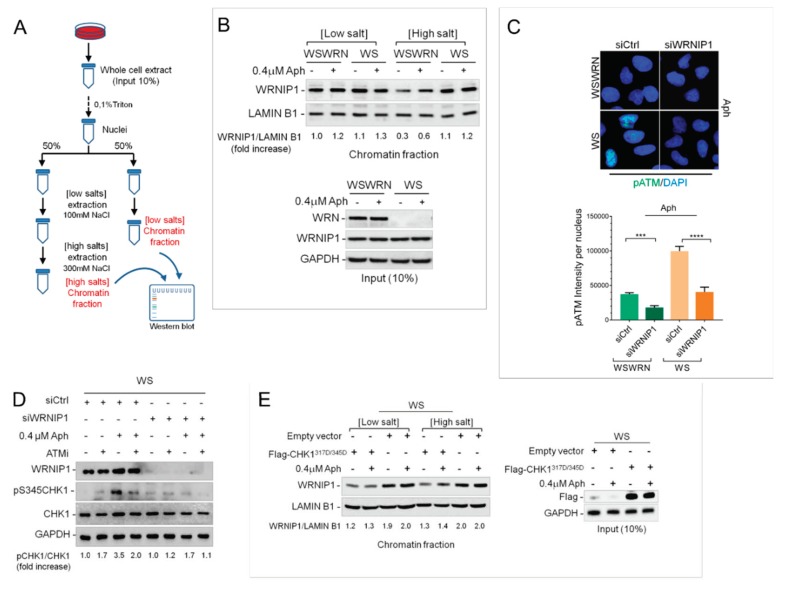
Figure 2.
WRNIP1 mediates the ATM-dependent CHK1 phosphorylation in WS cells. (A) Schematic representation of chromatin fractionation assay; (B) The membrane was probed with the indicated antibodies. The amount the chromatin-bound WRNIP1 is reported as a ratio of WRNIP1/LAMIN B1 normalized over the untreated control; (C) IF analysis of cells transfected with Green Fluorecent Protein (GFP) or WRNIP1 siRNA and stained for pATM (S1981). Bar graph shows pATM intensity per nucleus. Error bars represent standard error; (D) WB analysis of the presence of activated, i.e., phosphorylated, CHK1 assessed using S345 phospho-specific antibody (pS345) in WS cells depleted for WRNIP1 and treated with Aph. ATMi was added 1 h prior to Aph and used as a negative control. The membrane was probed with the indicated antibodies. The normalized ratio of the phosphorylated CHK1/total CHK1 is given. (E) WB analysis of chromatin binding of WRNIP1, performed as in (A) in WS cells transfected with empty vector or FLAG-tagged CHK1317/345D and treated with Aph. The membrane was probed with the indicated antibodies. The normalized ratio of the WRNIP1/LAMIN B1 signal (chromatin) is reported.

