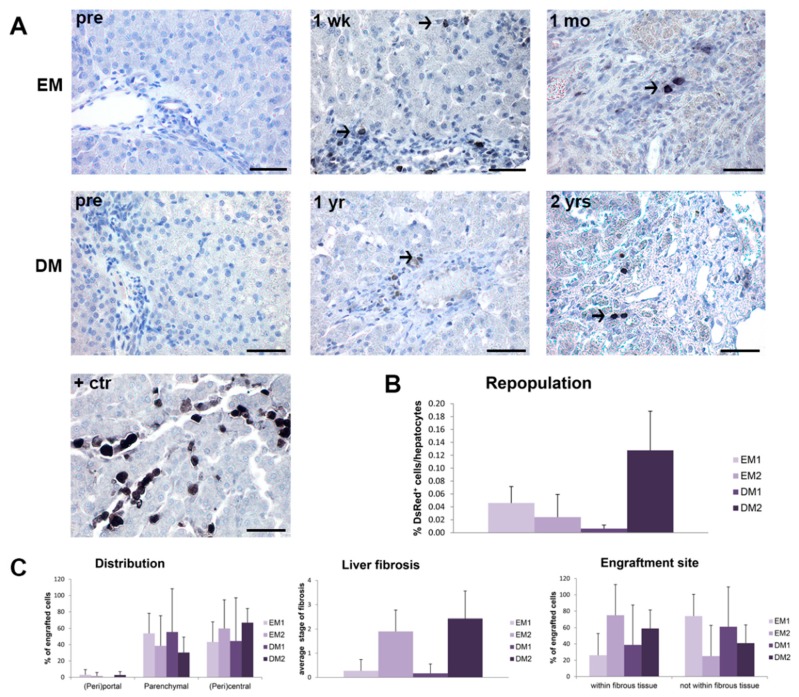
Figure 4.
Engraftment and long-term survival of organoid-derived liver cells after intraportal delivery. Four COMMD1-deficient dogs were transplanted via the portal vein and liver was sampled at various time points after transplantation for cell tracking purposes. (A) Representative images of immunohistochemical staining for DsRed in liver sections of undifferentiated (EM) or differentiated (DM) organoid-derived liver cells pre-transplantation (pre) and one week (1 wk: dog EM1), one month (1 mo: dog EM2), one year (1 yr: dog DM1) and two years (2 yrs: dog DM2) post-transplantation. Normal dog liver injected post-mortem with DsRed-transduced organoid-derived liver cells was used as positive control (+ctr). (B) Repopulation of liver with DsRed-positive cells expressed as percentage of total hepatocyte count. (C) Histologic distribution of transplanted cells and engraftment in either fibrous or non-fibrous tissue was determined and expressed as percentage of engrafted cells. Average stage of fibrosis was scored in liver sections after transplantation (2–10 sections per dog). Scale bars represent 50 µm.

