Figure 5.
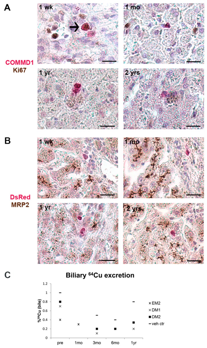
Proliferation and differentiation of transplanted cells in vivo. Double immunohistochemical staining was performed on liver sections post-transplantation to investigate the presence of proliferation marker Ki67 and differentiation marker MRP2 on transplanted cells. (A) Transplanted cells were sporadically positive for Ki67 (arrow), but only in sections one week after transplantation and not at later time points. (B) Transplanted cells did not show immunostaining for MRP2, whereas hepatocytes showed positive canalicular staining. 1 wk: one week (dog EM1); 1 mo: one month (dog EM2); 1 yr: one year (dog DM1); 2 yrs: two years (dog DM2) post- transplantation. Scale bars represent 20 µm. (C) Biliary copper excretion before (pre) and after transplantation indicates that the copper excretion remains low with all tested conditions. Pre: before transplantation; 1 mo: one month; 3 mo: three months; 6 mo: six months; 1 yr: one year after transplantation.

