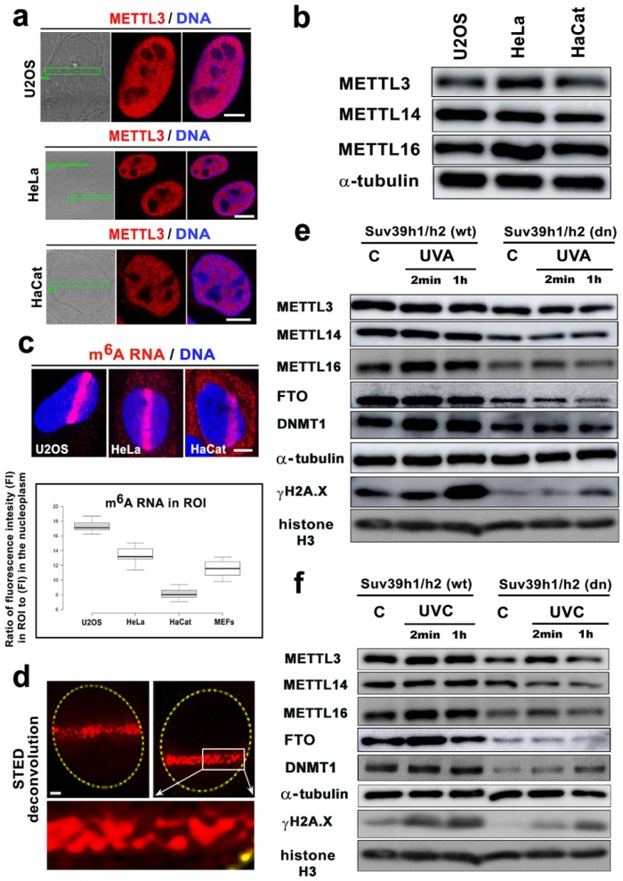
Figure 6.
The level of m6A RNAs in microirradiated chromatin was the highest in the U2OS osteosarcoma cells. (a) METTL3 enzyme was not changed in the microirradiated U2OS and HeLa tumor cells or the HaCaT keratinocytes. (b) Western blotting also showed that the levels of METTL3, METTL14, and METTL16 in U2OS, HeLa, and HaCaT cells were almost identical. (c) In comparison to the other cell types, U2OS cells were characterized by the most pronounced accumulation of m6A RNAs at DNA lesions. In HaCaT cells, the level of m6A RNAs at the CPD sites was the lowest compared to that of the other cell types studied (see box plot). Scale bars in panels a-c represent 8 µm. (d) STED microscopy showed the focal distribution of the m6A RNAs in the microirradiated regions. Scale bars represent 2 µm. Western blot analysis of the levels of METTL3, METTL14, METTL16, FTO, DNMT1, and γH2AX in Suv39h1/h2 wt and Suv39h1/h2 dn nonirradiated cells and cells exposed to (e) a UVA lamp and (f) a UVC lamp. Protein levels were normalized to the levels of α-tubulin, and the level of histone proteins was normalized to total histone H3.

