Figure 6.
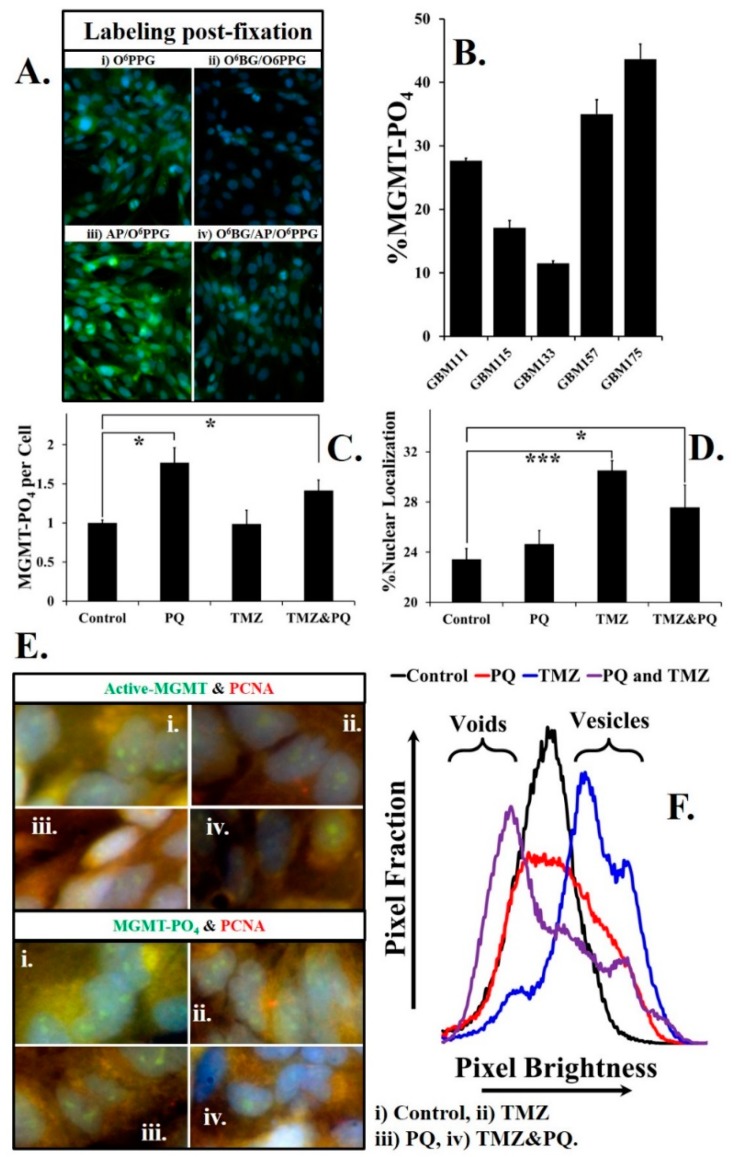
Phosphorylated MGMT in fixed cells can be labeled after reactivation by alkaline phosphatase. (A) (i) GBM157 cells labeled using O6PGG; (ii) treatment with MGMT inhibitor O6BG eliminates the O6PGG labeling of MGMT; (iii) pre-treatment of GBM157 cells with alkaline phosphatase (AP) increases MGMT labeling compared to control because of the conversion of phosphorylated MGMT into active MGMT; (iv) reactivation of phosphorylated (inactive) MGMT by AP in cells pre-treated with the MGMT inhibitor O6BG. (B) The fraction of phosphorylated (inactive) MGMT in different primary GBM cell lines show significant variations, and may reflect tumor heterogeneity; also see Supplementary Figure S3. (C) Statistically significant (t-test) increases in phosphorylated (inactive) MGMT levels occur upon treating GBM157 cells with either paraquat (PQ), or TMZ, or a combination of PQ and TMZ. (D) Statistically significant increases in phosphorylated (inactive) MGMT levels occur upon treating GBM157 cells with TMZ or a combination of PQ and TMZ. (E) Proliferating cell nuclear antigen (PCNA) levels were measured using a fluorescently labeled PCNA antibody. Active MGMT levels were measured using O6PGG/azido-PEG-FITC/click. Levels of phosphorylated (inactive) MGMT were measured by pre-treating GBM157 cells with O6BG followed by treatment with alkaline phosphatase and labeling using O6PGG/azido-PEG-FITC/click. (F) Analysis of the distribution of PCNA labeled pixels in the GBM157 nuclei. Treatment of GBM157 cells with PQ or TMZ or a combination of PQ and TMZ changes the line shape, indicating higher localization in nuclear vesicles in response to DNA damage.* p < 0.05, *** p < 0.001

