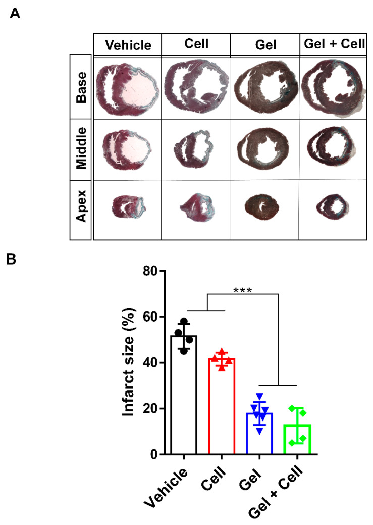
Figure 4.
(RADA)4-SDKP hydrogel diminished scar size. (A) Representative images of Masson’s trichrome (MT) stained heart sections at the apex, middle, and base areas for all groups. (B) At day 28, the infarct area (% of left ventricle (LV)) was decreased in the Gel and Gel + Cell groups in comparison with the Cell and Vehicle groups. All data are presented as mean ± standard deviation (n ≥ 4). *** p < 0.001.

