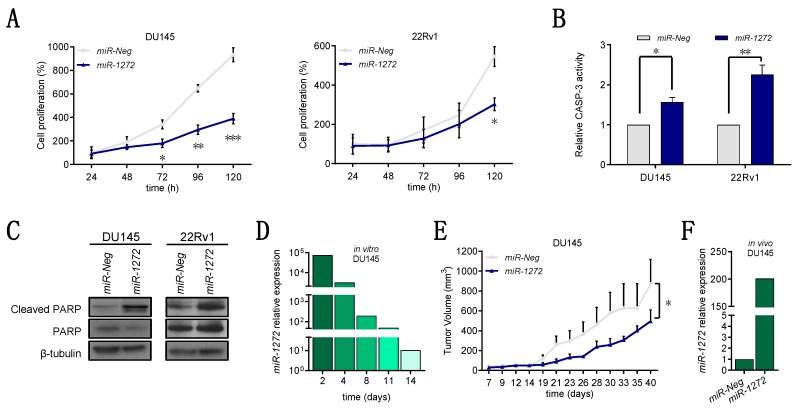
Figure 2.
miR-1272 reduces the growth of PCa cell lines both in vitro and in vivo. (A) Graphs reporting the growth of miR-Neg and miR-1272 transfected DU145 (top) and 22Rv1 (bottom) cells at different time points after transfection. Data are reported as percentage (mean ± SD as from 3 independent experiments) of cell number in each condition with respect to the number of miR-Neg cells collected at 24 h. (B) Bar plots reporting the relative catalytic activity of Caspase-3 (mean + SD from 3 independent experiments), an indicator of apoptosis induction, at 96 h after transfection of DU145 and 22Rv1 cells with miR-Neg and miR-1272. (C) Representative immunoblotting showing protein levels of cleaved and total PARP in miR-Neg and miR-1272 transfected DU145 (left) and 22Rv1 (right) cells. β-tubulin was used as endogenous control. (D) RT-qPCR showing relative expression of miR-1272 in miR-1272 transfected DU145 cells with respect to miR-Neg cells, at different time points after transfection. RNU48 was used as endogenous control. (E) Graph reporting the tumor growth volume (mean + SD) of xenografts originated from miR-Neg and miR-1272 transfected DU145 cells (10 mice per group), as measured with a Vernier caliper on the indicated days after cell injection into SCID mice. (F) RT-qPCR showing relative expression of miR-1272 in xenografts derived from miR-1272 transfected DU145 cells as compared to those derived from miR-Neg cells, at 23 days after injection into SCID mice. RNU48 was used as endogenous control. * p < 0.05, ** p < 0.01, *** p < 0.001, Student’s t-test.

