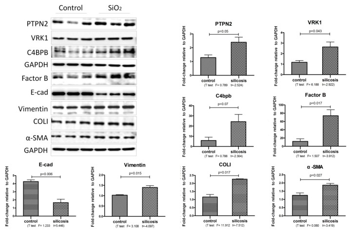
Figure 4.
The increased proteins expression in SiO2 stimulated MLE-12 cells. Western blot and corresponding densitometry data of the expression levels of PTPN2, factor B, VRK1, C4BPB, E-cad, Vimentin, Col I and α-SMA in MLE-12 cells on SiO2 (50 μg/mL) stimulated 24 h. Statistical analysis was performed using a t-test with SPSS20.

