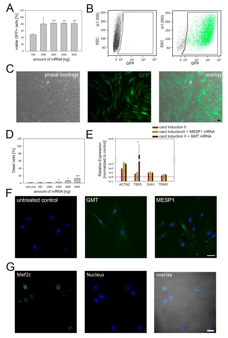
Figure 4.
mRNA-based cardiac programming of adMSC. (A) Concentration-dependent expression of transfected mRNAs was evaluated with mRNA coding GFP. The quantative flow cytometry analysis demonstrated maximum transfection efficiency of ~80% when ≤ 1000 ng mRNA were applied. (B) Representative scatterplots of control cells (left) and cells transfected with GFP mRNA (right). (C) Corresponding microscopy images of cells expressing GFP following mRNA treatment. (D) Cytotoxic effects were only induced when mRNA amounts higher than 1000 ng were used for transfection. (E) Compared to untreated control cells, higher gene expression levels of selected cardiac markers were detected for all reprogramming conditions, in particular for α-actinin. (F) Immunolabeling of cells using anti α-actinin antibody results in a faint fluorescence signal in cells transfected with MESP1 and GATA4, MEF2C, and TBX5 (GMT) mRNAs, Scale Bar: 25 µm. (G) Moreover, GMT treated cells also demonstrated protein expression of MEF2C, an early cardiac transcription factor. Flow cytometry and qRT-PCR data are shown as mean ± SEM, obtained from three different donors. Statistical analysis was performed using one-way ANOVA. * p ≤ 0.5, ** p ≤ 0.05, *** p ≤ 0.001.

