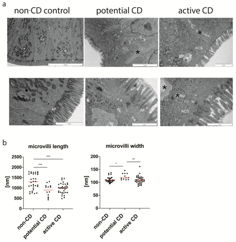
Figure 3.
Ultrastructure of small intestine enterocytes from the non-CD control group, patients with potential and active CD (a). Cross-sections of non-CD enterocytes with anchoring filaments (AF), endosomes (e) and no intracellular dilatations between cells present in the control group. Enterocytes in potential and active CD exhibit numerous endosomes (e) and tubules of an apical canicular system (ACS), and dilated intercellular spaces (*). The length and width of the brush border microvilli (b). Measurements were done with the use of the morphometric iTEM program (Olympus) in 10 selected epithelial areas at a magnification of ×60,000, and at least 3 values/patient were obtained. All measurements are presented. Statistical analysis was performed with the use of one-way ANOVA with Tukey correction for multiple comparisons. * p < 0.05, ** p ≤ 0.01, *** p ≤ 0.001.

