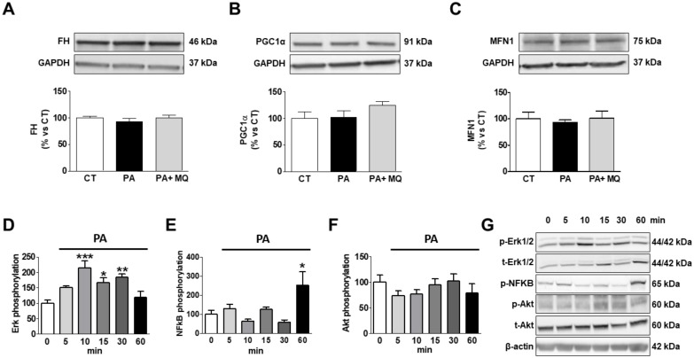
Figure 8.
Effects of antioxidant MitoQ (MQ) on protein levels in palmitic acid (PA)-treated H9c2 cells. Protein levels of (A) fumarate hydratase (FH), (B) peroxisome proliferator-activated receptor gamma coactivator 1-alpha (PGC-1α), (C) mitofusin 1 (MFN1) in cardiac myoblasts treated for 24 h with PA (200 µM) in the presence of absence of the mitochondrial antioxidant MitoQ (MQ; 5 nM). (D) Phosphorylated protein of extracellular signal-regulated kinases (pErk1/2), (E) phosphorylated protein of nuclear factor-κB p65 (pNF-κB), and (F) phosphorylated protein kinase B (pAkt) in cardiac myoblasts treated with PA (200 µM) at different times. (G) Representative images blots for phosphorylation of intracellular signaling pathways. Bar graphs represent the mean ± SEM of four to six assays normalized to glyceraldehyde-3-phosphate dehydrogenase (A–C), β-actin, (E) or total Erk1/2 (D) and total Akt (F). * p < 0.05; ** p < 0.01; *** p < 0.001 vs. control conditions.

