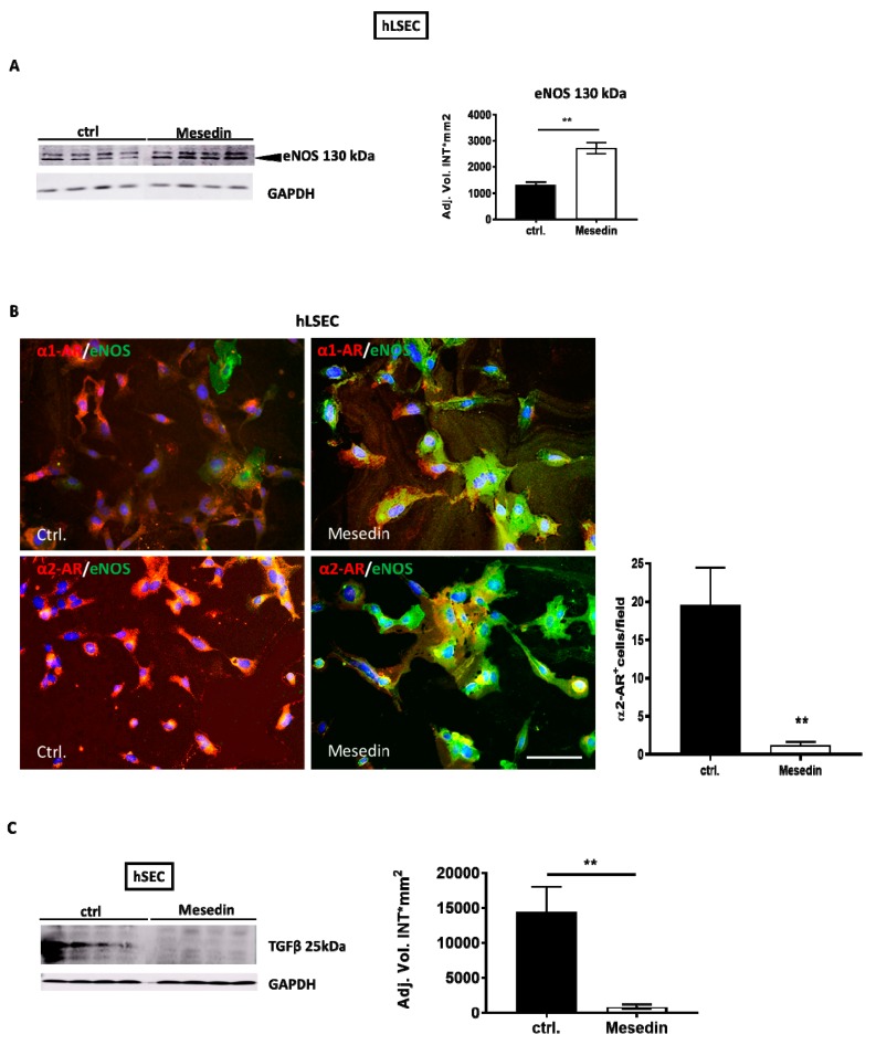
Figure 6.
Impact of mesedin on the permeability of human liver sinusoidal endothelial cells (hLSECs). (A) Western blot and densitometric analysis of eNOS in hLSECs with and without (ctrl.) mesedin, n = 4. (B) Immunofluorescence analysis of α1-AR vs α2-AR (red) and eNOS (green) in hLSECs. Cell nuclei were stained with DAPI. Scale bar corresponds to 100 µm. Quantification of α2-AR-positive cells in hLSECs (n = 6, p < 0.01 (**), Student’s t-test, data shown as the mean ± SEM). (C) Western blot and densitometric analysis of TGF-β in hLSECs 48 h after incubation with and without (ctrl.) mesedin. Student’s t-test, p < 0.01 (**).

