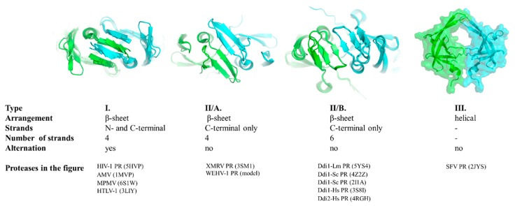
Figure 3.
Classification of dimer interfaces. Dimer interfaces are represented according to selected structures, PDBIDs are shown for each protease. Homology model is shown for WEHV-1 PR. Structure of homodimeric SFV PR was proposed by aligning the monomers to a homodimeric HIV-2 PR structure (PDBID: 1HII). Subunits are colored by green and cyan.

