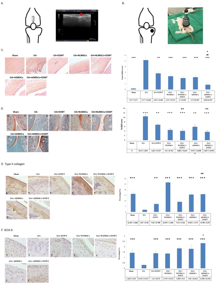
Figure 2.
Treatments, histological analysis of synovium membrane, and Osteoarthritis Research Society International (OARSI) scores of the knee osteoarthritis (OA). (A) Sketch of the knee outlining the location of injection of WJMSCs or ADMSCs in the rats. In the left, ultrasound guidance (Toshiba Medical Systems Corporation, Tokyo, Japan) was used for localization, and WJMSCs or ADMSCs were injected into the knees of the rats. The red arrow indicates the needle of the injector. (B) Knee sketch outlining the locations (black dots) of shockwave application in the different groups of animals. The left picture showed that extracorporeal shockwave therapy (ESWT) was applied to the knees of the rats; n = 6. (C) The synovium membrane was displayed by hematoxylin and eosin staining at magnifications of 200×. The left panel shows synovitis scores were measured in each group. (D) Microphotographs of cartilage and subchondral bone showed changes in cartilage damage in the OA group and protection in the treatment groups by safranin O staining. (E) Type II collagen (×200 magnification). (F) SOX-9 (×400 magnification). Scale bar represents 200 μm; T indicates the tibia; F indicates the femur. ** p < 0.01, and *** p < 0.001 were compared with OA; # p < 0.05, ## p < 0.01, and ### p < 0.001 were compared among treatment groups. ✦ p < 0.05 was compared ESWT combined with ADMSCs or WJMSCs each other. n = 6 for each group.

